 SDS
SDS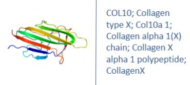
Other Names
Human Collagen Type X ELISA kit
Collagen type X; Col10a 1; Collagen alpha 1(X) chain; Collagen type X alpha 1 (Schmid metaphyseal chondrodysplasia); Collagen type X alpha 1; Collagen X alpha 1 polypeptide; CollagenX; Procollagen type X alpha 1; Schmid metaphyseal chondrodysplasia; Collagen alpha-1(X) chain; collagen alpha-1(X) chain precursor; Schmid metaphyseal chondrodysplasia; collagen X, alpha-1 polypeptide
Research Area
Signal transduction
Background
This gene encodes the alpha chain of type X collagen, a short chain collagen expressed by hypertrophic chondrocytes during endochondral ossification. Unlike type VIII collagen, the other short chain collagen, type X collagen is a homotrimer. Mutations in this gene are associated with Schmid type metaphyseal chondrodysplasia (SMCD) and Japanese type spondylometaphyseal dysplasia (SMD).
|
Product Name |
COL10 ELISA |
|
Species |
Human |
|
Product Size |
96/48 Tests |
|
Concentration |
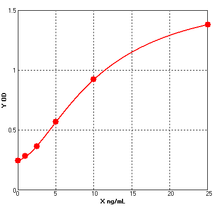
1.0-25 ng/mL |
|
Sensitivity |
0.09 ng/mL |
|
Principle |
Sandwich ELISA |
|
Sample Volume |
50 ul |
|
Assay Time |
90 minutes |
|
Platform |
Microplate Reader |
|
Conjugate |
HRP |
|
Detection Method |
Colorimetric |
|
Storage |
2-8°C |
|
|
For reference use only.
Four parameter Logisticcurve regression
Formular: y = (A - D) / [1 + (x/C)^B] + D
A = 1.64480
B = -1.65539
C = 10.42106
D = 0.24982
r^2 = 0.99993
|
1. Protocols for ELISA
![]() 1) Direct ELISA
1) Direct ELISA
![]() 2) Direct ELISA Using Fluorescent Substrate
2) Direct ELISA Using Fluorescent Substrate
![]() 3) Indirect ELISA
3) Indirect ELISA
![]() 4) Sandwich ELISA
4) Sandwich ELISA
2. Protocols for IHC ICC
![]() 1) Determining if the antibody binds only phosphorylated protein (WB or IHC)
1) Determining if the antibody binds only phosphorylated protein (WB or IHC)
![]() 2) Double immunofluorescence-sequential protocol
2) Double immunofluorescence-sequential protocol
![]() 3) Double immunofluorescence-simultaneous protocol
3) Double immunofluorescence-simultaneous protocol
![]() 4) Fixation and Permeabilization In IHC ICC
4) Fixation and Permeabilization In IHC ICC
![]() 5) Glycol Methalacrylate Acrylic Resin Embedding For IHC
5) Glycol Methalacrylate Acrylic Resin Embedding For IHC
![]() 9) Immunohistochemistry (IHC-Fr) - Frozen Sections
9) Immunohistochemistry (IHC-Fr) - Frozen Sections
3. Protocols for WB
![]() 4) S-100 Mitochondrial Fractionation
4) S-100 Mitochondrial Fractionation
![]() 5) Stripping for Reprobing Western Blots
5) Stripping for Reprobing Western Blots
![]() 7) Western Blotting - A Beginner's Guide
7) Western Blotting - A Beginner's Guide
![]() 8) Western Blotting of Phospho-Proteins
8) Western Blotting of Phospho-Proteins
![]() 9) Western Blotting Using Antibodies Against Histone Proteins
9) Western Blotting Using Antibodies Against Histone Proteins
4. Protocols for IP
![]() 2) Using IgM antibodies for IP
2) Using IgM antibodies for IP
5. Protocols for FACS
![]() 1) Direct Staining Protocol (Cell Surface Staining)
1) Direct Staining Protocol (Cell Surface Staining)
![]() 3) Flow Cytometry Whole Blood Samples-Red Blood Cell Lysis
3) Flow Cytometry Whole Blood Samples-Red Blood Cell Lysis
![]() 4) Indirect Staining Protocol (Cell Surface Staining)
4) Indirect Staining Protocol (Cell Surface Staining)
![]() 6) Recommended Controls for FACS
6) Recommended Controls for FACS
6. Protocols for ELISPOT
![]() 1) ELISPOT
1) ELISPOT


