Other Names
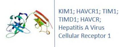
HAVCR1; TIM1; TIMD1; HAVCR; Hepatitis A Virus Cellular Receptor 1; T Cell Immunoglobulin And Mucin Domain-Containing Protein 1; T-cell immunoglobulin mucin receptor 1
Research Area
Immunology, Microbiology
Background
Traditional blood and urine markers for the diagnosis of various renal diseases are insensitive and nonspecific. Kidney Injury Molecule-1 (KIM-1) is a type 1 transmembrane protein, with an immunoglobulin and mucin domain, whose expression is markedly up-regulated in the proximal tubule in the post-ischemic rat kidney. The ectodomain of KIM-1 is shed from cells. The current studies were carried out to evaluate whether KIM-1 is present in human acute renal failure and might serve as a urinary marker of acute renal tubular injury.
|
Product Name |
KIM1ELISA |
|
Species |
Monkey |
|
Product Size |
96/48 Tests |
|
Concentration |
1.0-25 ng/ml |
|
Sensitivity |
0.09 ng/ml |
|
Principle |
Competitive ELISA |
|
Sample Volume |
100 ul |
|
Assay Time |
90 minutes |
|
Platform |
Microplate Reader |
|
Conjugate |
HRP |
|
Detection Method |
Colorimetric |
|
Storage |
2-8°C |
|
|
For Research use only
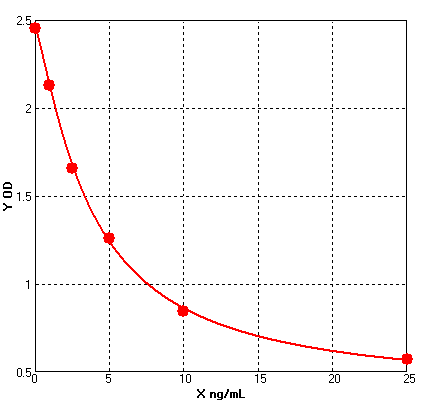
Formular: y = (A - D) / [1 + (x/C)^B] + D
A = 2.45541 B = 1.23695 C = 3.76072 D = 0.38392 r^2 = 0.99960 |
1. Protocols for ELISA
![]() 1) Direct ELISA
1) Direct ELISA
![]() 2) Direct ELISA Using Fluorescent Substrate
2) Direct ELISA Using Fluorescent Substrate
![]() 3) Indirect ELISA
3) Indirect ELISA
![]() 4) Sandwich ELISA
4) Sandwich ELISA
2. Protocols for IHC ICC
![]() 1) Determining if the antibody binds only phosphorylated protein (WB or IHC)
1) Determining if the antibody binds only phosphorylated protein (WB or IHC)
![]() 2) Double immunofluorescence-sequential protocol
2) Double immunofluorescence-sequential protocol
![]() 3) Double immunofluorescence-simultaneous protocol
3) Double immunofluorescence-simultaneous protocol
![]() 4) Fixation and Permeabilization In IHC ICC
4) Fixation and Permeabilization In IHC ICC
![]() 5) Glycol Methalacrylate Acrylic Resin Embedding For IHC
5) Glycol Methalacrylate Acrylic Resin Embedding For IHC
![]() 9) Immunohistochemistry (IHC-Fr) - Frozen Sections
9) Immunohistochemistry (IHC-Fr) - Frozen Sections
3. Protocols for WB
![]() 4) S-100 Mitochondrial Fractionation
4) S-100 Mitochondrial Fractionation
![]() 5) Stripping for Reprobing Western Blots
5) Stripping for Reprobing Western Blots
![]() 7) Western Blotting - A Beginner's Guide
7) Western Blotting - A Beginner's Guide
![]() 8) Western Blotting of Phospho-Proteins
8) Western Blotting of Phospho-Proteins
![]() 9) Western Blotting Using Antibodies Against Histone Proteins
9) Western Blotting Using Antibodies Against Histone Proteins
4. Protocols for IP
![]() 2) Using IgM antibodies for IP
2) Using IgM antibodies for IP
5. Protocols for FACS
![]() 1) Direct Staining Protocol (Cell Surface Staining)
1) Direct Staining Protocol (Cell Surface Staining)
![]() 3) Flow Cytometry Whole Blood Samples-Red Blood Cell Lysis
3) Flow Cytometry Whole Blood Samples-Red Blood Cell Lysis
![]() 4) Indirect Staining Protocol (Cell Surface Staining)
4) Indirect Staining Protocol (Cell Surface Staining)
![]() 6) Recommended Controls for FACS
6) Recommended Controls for FACS
6. Protocols for ELISPOT
![]() 1) ELISPOT
1) ELISPOT

 SDS
SDS


