 SDS
SDS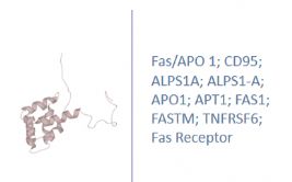
Other Names
CD95; ALPS1A; ALPS1-A; APO1; APT1; FAS1; FASTM; TNFRSF6; Fas Receptor; TNF Receptor Superfamily Member 6; Tumor Necrosis Factor Receptor Superfamily Member 6
Research Area
Cell Biology, Immunology
Background
FasL, the physiological agonist for Fas, is also a transmembrane protein with homology to the TNF family in its extracellular domain. FasL is expressed primarily by activated T lymphocytes and by cells of the small intestine and lung. Mice with mutations in either Fas or FasL exhibit accumulation of activated lymphocytes and classical autoimmune symptoms, suggesting that a major function of Fas-mediated apoptosis is the elimination of activated immune cells from the peripheral circulation. Similarly, humans with autoimmune lymphoproliferative syndrome have mutations in Fas. Fas and FasL have been observed as soluble molecules in addition to their membraneassociated forms, suggesting additional complexity to regulation of this apoptotic mechanism. Soluble Fas (sFas) arises from alternatively spliced mRNA, leading to proteins with deletion or disruption of the single membrane-spanning domain. Five alternatively spliced Fas mRNAs have been described, each protein detected in the supernate of cultures of peripheral blood mononuclear cells or certain tumor cell lines. Each sFas inhibited apoptosis induced by FasL, and tumor-cell lines resistant to anti-Fas were shown to produce alternatively spliced Fas, thereby making them less sensitive to FasL. In addition, plasma Fas can arise by exfoliation of membrane vesicles, which also inhibit FasL-induced apoptosis. Serum Fas has been reported to be elevated in cancer patients , possibly originating in the tumor cell itself), and in autoimmune diseases
|
Product Name |
Fas/APO 1 ELISA |
|
Species |
Rabbit |
|
Product Size |
96/48 Tests |
|
Concentration |
250-5000pg/mL |
|
Sensitivity |
21.5 pg/mL |
|
Principle |
Competitive ELISA |
|
Sample Volume |
100 ul |
|
Assay Time |
90 minutes |
|
Platform |
Microplate Reader |
|
Conjugate |
HRP |
|
Detection Method |
Colorimetric |
|
Storage |
2-8°C |
|
|
For Research use only
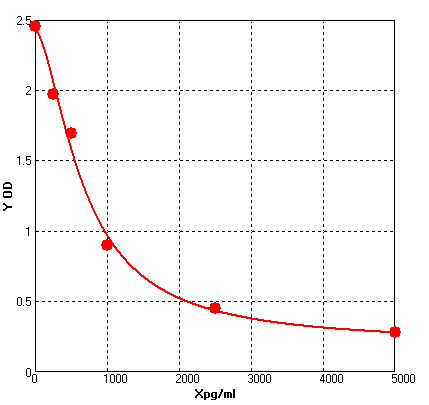
Four parameter Logisticcurve regression
A = 1.77782 B = 1.62110 C = 303.27773 D = 0.04416 r^2 = 0.99777 |
1. Protocols for ELISA
![]() 1) Direct ELISA
1) Direct ELISA
![]() 2) Direct ELISA Using Fluorescent Substrate
2) Direct ELISA Using Fluorescent Substrate
![]() 3) Indirect ELISA
3) Indirect ELISA
![]() 4) Sandwich ELISA
4) Sandwich ELISA
2. Protocols for IHC ICC
![]() 1) Determining if the antibody binds only phosphorylated protein (WB or IHC)
1) Determining if the antibody binds only phosphorylated protein (WB or IHC)
![]() 2) Double immunofluorescence-sequential protocol
2) Double immunofluorescence-sequential protocol
![]() 3) Double immunofluorescence-simultaneous protocol
3) Double immunofluorescence-simultaneous protocol
![]() 4) Fixation and Permeabilization In IHC ICC
4) Fixation and Permeabilization In IHC ICC
![]() 5) Glycol Methalacrylate Acrylic Resin Embedding For IHC
5) Glycol Methalacrylate Acrylic Resin Embedding For IHC
![]() 9) Immunohistochemistry (IHC-Fr) - Frozen Sections
9) Immunohistochemistry (IHC-Fr) - Frozen Sections
3. Protocols for WB
![]() 4) S-100 Mitochondrial Fractionation
4) S-100 Mitochondrial Fractionation
![]() 5) Stripping for Reprobing Western Blots
5) Stripping for Reprobing Western Blots
![]() 7) Western Blotting - A Beginner's Guide
7) Western Blotting - A Beginner's Guide
![]() 8) Western Blotting of Phospho-Proteins
8) Western Blotting of Phospho-Proteins
![]() 9) Western Blotting Using Antibodies Against Histone Proteins
9) Western Blotting Using Antibodies Against Histone Proteins
4. Protocols for IP
![]() 2) Using IgM antibodies for IP
2) Using IgM antibodies for IP
5. Protocols for FACS
![]() 1) Direct Staining Protocol (Cell Surface Staining)
1) Direct Staining Protocol (Cell Surface Staining)
![]() 3) Flow Cytometry Whole Blood Samples-Red Blood Cell Lysis
3) Flow Cytometry Whole Blood Samples-Red Blood Cell Lysis
![]() 4) Indirect Staining Protocol (Cell Surface Staining)
4) Indirect Staining Protocol (Cell Surface Staining)
![]() 6) Recommended Controls for FACS
6) Recommended Controls for FACS
6. Protocols for ELISPOT
![]() 1) ELISPOT
1) ELISPOT


