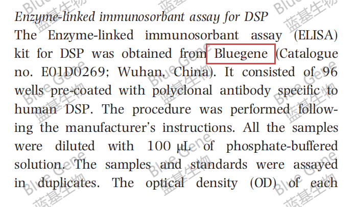
Human DSP Elisa kit citation
文件下载

2017-10-26940
Abstract
Kumar V, Logani A, Shah N. Dentine sialoprotein expression in gingival crevicular fluid during trauma-induced root resorption. International Endodontic Journal.
Aim
To detect and quantify dentine sialoprotein (DSP) in the gingival crevicular fluid (GCF) of luxated teeth.
Methodology
Eighteen subjects were enroled and distributed as follows. Group I (n = 6, positive control): subjects with primary second molar teeth undergoing physiological root resorption. Group II (n = 6,negative control): subjects with permanent mature maxillary central incisors. Subjects with a recent history (<1 week) of luxation injury were included in group III (n = 6, test group) and standardized digital radiographs with a superimposed mesh gauge were
exposed at various time intervals. Percentage of radiographic root resorption (%RRR) was calculated. GCF was collected using microcapillary pipettes. DSP in the GCF was quantified using enzyme-linked immunosorbant assay. Group III was subjected to Spearman’s rank test to establish the correlation between the concentration of DSP and %RRR at 6 weeks, 3 and 6 months.
Results
Quantifiable amounts of DSP were released in the GCF of subjects in Group I and III. However,the protein was not detected in Group II. Detectable quantities of DSP were observed in the GCF of luxated teeth before any radiographic vidence of root resorption (base line radiograph). A positive correlation was established at 6 weeks (r = 0.795), 3 (r = 0.755) and 6 month (r = 0.837) between the release of DSP and %RRR (P < 0.05).